
Overcoming the optical resolution limit with microspheres
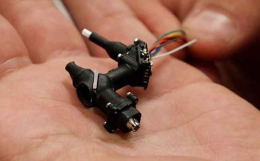
New approach enables interferometric topography measurements.
New approach enables interferometric topography measurements.
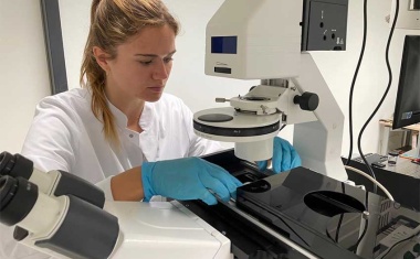
With a multimodal microscope, the joint project PriMe wants to detect bacterial infestation using fast, marker-free, and contactless imaging.
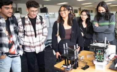
Cloud-controlled microscope brings complex biology education to students worldwide.

A new type of microscope can take 3D images of cells while working in a natural environment.
New technology could enable point-of-care skin cancer diagnosis.
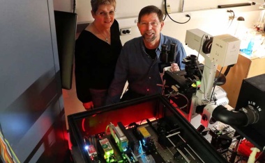
The development of a microscope for the gentle 3D imaging of living cells was honored with the Federal President's Award for Technology and Innovation

PicoQuant has created a system that is easier to use without any compromise on sensitivity. It makes these systems accessible for users who do not enjoy the support of a trained physicist.
New kind of label-free deep-tissue imaging retrieves the fine neural network of a mouse brain.
Innovative microscope makes it easier and faster to diagnose cancer.
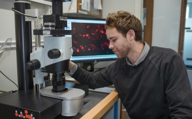
Detecting much more subtle biological events using a neural network.
A frequency-domain method possesses great potential to improve STED microscopy.
The UCLA creators of a miniature microscope that can be mounted on the heads of lab animals to provide an invaluable view into the brain’s inner workings have received a 4 million dollar grant from the National Institutes of Health to develop next-generation versions of their “miniscope.”

German researchers combine two techniques to achieve isotropic super-resolution imaging.
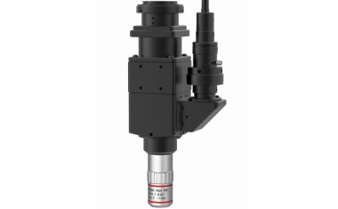
A metrology manufacturer has equipped a video microscope for research and industrial quality assurance with a larger field of view and higher illumination quality.
The realization of light-based aberration corrector will significantly reduce installation costs for electron microscopes with atomic resolution.

Resolving nanostructures smaller than the diffraction limit of light without any dyes or labels.
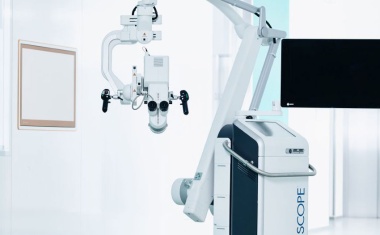
The new collaborative project ‘BetterView’ is working on a special surgical microscope in order to detect cholesteatomas – an aggressive form of chronic otitis media – and bacterial biofilms and to remove them safely.
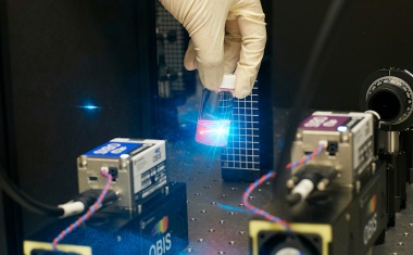
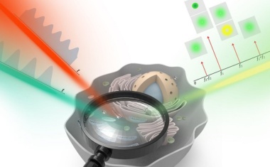
New device can measure the very detailed dynamics of how molecules move and rotate inside liquid system.
Oregon Health & Science University researchers are developing an entirely new approach to scientific imaging to learn how the seemingly random movements of molecules are actually a well-orchestrated operation.

With their new instrument, University of California researchers achieve a large field of view up to 25 square millimeters to provide subcellular resolution of multiple areas of the brain.

An electron beam was steered through the optical near field of a photonic circuit, to allow the electrons to interact with the enhanced light.
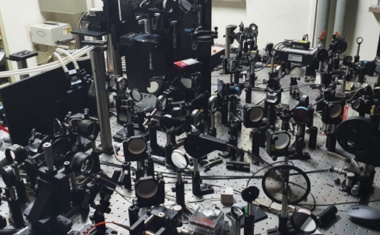
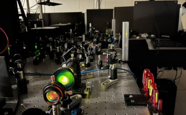
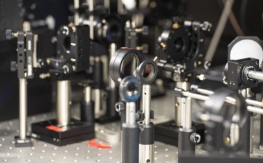
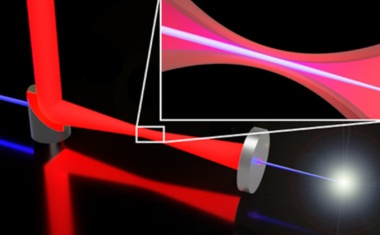
A new microscope uses interference patterns of entangled photons and very low intensity levels of light to image large areas of samples with a high sensitivity and resolution.

The Annual General Meeting of Carl Zeiss AG appointed Dr. Michael Bolle as a new member of the Supervisory Board. He is to be elected Chairman of the the Board in January 2022.

Zeiss achieved record sales and operating profit in fiscal year 2021 each. In addition, all divisions grew at double-digit rates. Group revenues, as a result, rose by 20 percent to EUR 7.5 billion and Ebit by 60 percent to EUR 1.48 billion, both compared to the previous year. Order intake grew to 8.97 billion euros, an increase of around 32 percent.
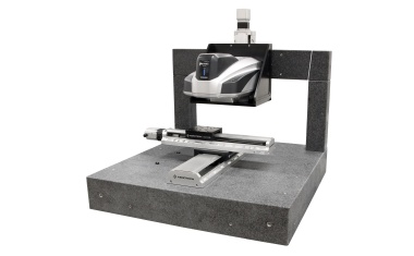
Peak Metrology, solution provider for the surface metrology sector, is now cooperating with IDC Microinspection. The partnership combines Peak Metrology's instrument and hardware capabilities with IDC MicroInspection's process knowledge and application software. As a result, users of digital microscopes benefit from new possibilities such as larger measurement volumes and greater automation in image acquisition.

A multinational team from the Institut Pasteur in Paris, the Francis Crick Institute in London and PoL scientists in Dresden will develop solutions for distributed, accelerated image analysis that is accessible to end-users without programming experience.
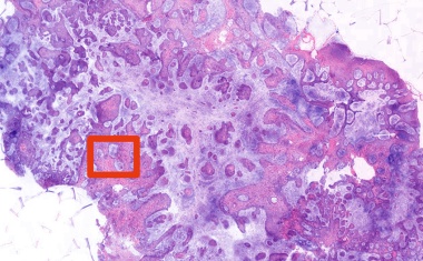
New method can be used to reveal previously unrecognised alterations in the pancreas, but it can also be used to study other human organs and diseases.
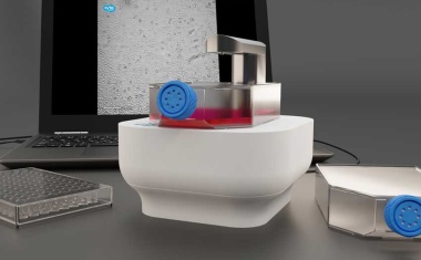
CytoSmart Technologies has launched a new brightfield live-cell imaging system – the Lux3 BR. It is a small brightfield microscope, equipped with a high-quality 6.4 MP CMOS camera.

The Zeiss Group closed fiscal year 2019/20 (balance sheet date: September 30, 2020) with a slight decline in revenue and operating profit.