Seeing through blood with a microscope
The new collaborative project ‘BetterView’ is working on a special surgical microscope in order to detect cholesteatomas – an aggressive form of chronic otitis media – and bacterial biofilms and to remove them safely.
The project’s SWIR microscope system uses short-wave infrared light. The aim is to illuminate blood, bacterial biofilms, cartilage, and soft tissue; display them spatially; and make them distinguishable from each other. The research is coordinated by the medical technology company Munich Surgical Imaging. The seven partner institutions cooperating in the project include Bielefeld University and Klinikum Bielefeld, one of the hospitals forming the University Hospital OWL. A total of 4.1 million euro will be spent on the project. The Federal Ministry of Education and Research is funding the new research.
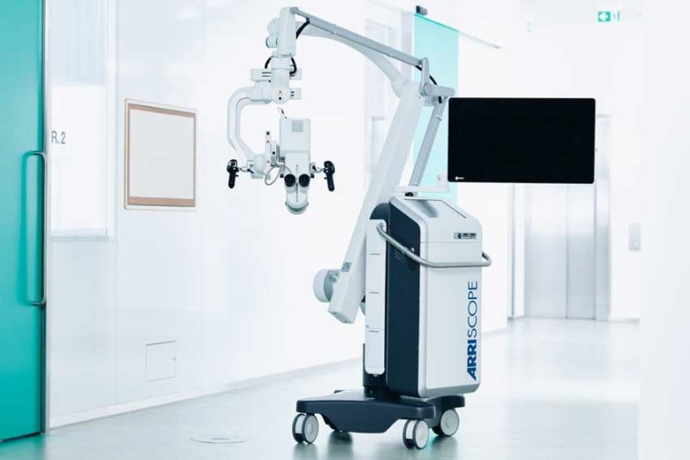

“An advanced generation of image sensors now makes it possible to equip surgical microscopes with a new function: to process and display images in the short-wave infrared light spectrum in real time,” says Thomas Huser from the faculty of physics at Bielefeld University. The professor is a specialist in biomedical photonics, which deals with the development of novel microscopy methods. Together with his team, he is constructing and using high-resolution microscopes while developing the software for image processing.
Microscopes with sensors such as the SWIR surgical microscope first have to analyze and process the recorded image signal automatically.
So that the surgical microscope can display the short-wave infrared signals, Huser and his team are developing their own software that filters out light outside the short-wave infrared spectrum and calculates a three-dimensional view of the image. “In addition, the software needs to produce color contrasts. Such colored markings make it easy to distinguish between, for example, nerves and soft tissue,” Huser explains. The software has to display the video image in real time so that surgeons in the operating theater can work precisely and face no delay in seeing what their intervention is doing to the surgical field.
In order to test the SWIR surgical microscope in practice, the project will initially use it to treat cholesteatoma – a chronic pus-producing inflammation of the middle ear. The microscope will be tested at the University Hospital OWL’s department of otorhinolaryngology, head, and neck surgery at the Klinikum Bielefeld. The clinic performs the most cholesteatoma operations nationwide – 650 procedures annually.
Compared to conventional microscopes, the future SWIR microscope will also be able to see through soft tissue. This will make it possible to examine optically hidden areas as well. Then, surgeons will be able to see whether bone material in the inner ear has been colonized or damaged by bacteria. In addition, the microscope should increase patient safety. If surgeons can see and distinguish the inner ear precisely, there is less risk of damaging sensitive structures such as the facial nerve or the labyrinths of the inner ear.
Alongside Bielefeld University and Klinikum Bielefeld, other members of the cooperation project are the Helmholtz Pioneer Campus at Helmholtz Zentrum München, Leibniz University Hannover, the camera system manufacturer PCO, and the laser manufacturer Omicron-Laserage Laserprodukte.
Company
Munich Surgical Imaging GmbHTuerkenstrasse 89
80799 Munich
Germany


most read

The market for humanoid robots is growing rapidly
Global production of humanoid robots rose to over 20,000 units in 2025, a tenfold increase compared to 2024.
Heitec takes over Artschwager + Kohl
Through the acquisition, the company aims to expand its range of warehouse logistics solutions and open up new market segments.

5 robotics trends for 2026
The International Federation of Robotics reports on the five most important trends for the robotics industry in 2026.
Siemens takes over Canopus AI
This expansion of the Siemens EDA software portfolio is designed to help chip manufacturers improve precision and efficiency in wafer and mask inspection processes.
Sick and Innok Robotics intensify partnership in the field of outdoor robotics
Both companies are now working on technological developments.