Real-time imaging of blood flow
New Mikrotron camera for life sciences and clinical medicine applications.
Visualization of myocardial blood flow is crucial for studying the pathogenesis of coronary heart disease. In the quality control of surgeries, such as coronary artery bypass grafting, there is also an urgent need to constantly image myocardial blood flow perfusion. Current techniques for monitoring myocardial blood flow are Transit Time Flow Measurement (TTFM) and Positron Emission Tomography-computed Tomography (PET). Although both are widely used, the processes either fail in sufficient spatial sampling or cannot perform dynamic real-time imaging.
Laser Speckle Contrast Imaging (LSCI), a less-used technique, is a real-time wide-field technology that can offer a powerful means to assess tissue blood flow in both human and animal tissues throughout a wide dynamic range (WDR). The full-field vision and high spatiotemporal resolution of LSCI is drawing interest from the scientific and medical communities. Scientists from China's Capital Medical University, Technical Institute of Physics and Chemistry, and University of Chinese Academy of Sciences have shown via animal experiments that the LSCI technique is capable of measuring velocities over a wide dynamic range in real-time. The imaging system documented the spatiotemporal evolution of myocardial perfusion in coronary artery bypass grafting, an advancement that could be of great benefit for future research in the life sciences and clinical medicine. All experiments were carried out in accordance with the guidelines for the humane care of animals.
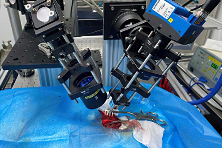
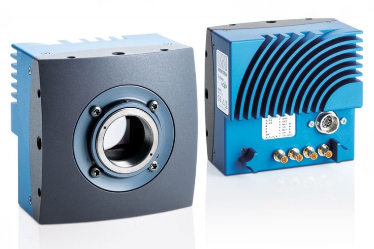
The experimental setup consisted of a 785 nanometer wavelength laser diode placed in a mount with a thermoelectric cooling stage. Before the samples were illuminated, a light pipe homogenized the laser beam. The imaging system itself was composed of a Mikrotron EoSens 4CXP CoaXPress CMOS camera set to acquire images at 2336 × 1728 resolution with 563 frames per second, along with a bandpass filter, polarizer, tube lens, plus an objective lens. The Mikrotron camera's four CXP-6 channels were connected to a single CXP frame grabber via coaxial cables transmitting at a total of 25 Gigabit per second. Using the camera, the WDR-LSCI method was successfully demonstrated to measure a high blood flow rate with superior performance when compared to TTFM. The new system showed higher sensitivity and the ability to obtain a wide-field, high-precision blood flow rate map in the non-contact way while imaging in vivo.
SVS-Vistek is under the TKH Vision umbrella in the TKH Group. Mikrotron EoSens CoaXPress cameras are available in either CXP6 or CXP12 interfaces in resolutions as high as 21 megapixels. Using four parallel lanes, data rates of up to 50 Gigabit per second are possible with the CXP12 designs in both monochrome and color. (Source: SVS-Vistek)
Link: Beijing Anzhen Hospital, Capital Medical U., Beijing, China • Mikrotron, SVS-Vistek GmbH, Gilching, Germany


most read

"Solutions and Products for Defense" Technology Day Showcases the Latest Innovations for Mission-Critical Applications
The free online event is now available as a recording at no cost and is aimed at users and integrators in the field of machine vision.

TPL Vision joins EMVA
The company is looking forward to working together and sharing knowledge within the industry
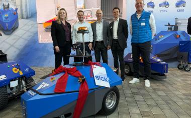
Sick and Innok Robotics intensify partnership in the field of outdoor robotics
Both companies are now working on technological developments.
Sick increases sales in 2025
In the 2025 financial year, the company increased its turnover to 1.85 billion euros, an increase of 6.5% compared to the previous year.
Heitec takes over Artschwager + Kohl
Through the acquisition, the company aims to expand its range of warehouse logistics solutions and open up new market segments.