Wide high-speed imaging with super-resolution
Microscope based on fiberoptic components designed for studying cellular effects of drug combinations.
Researchers have developed a fluorescence microscope that uses structured illumination for fast super-resolution imaging over a wide field of view. The new microscope was designed to image multiple living cells simultaneously with a very high resolution to study the effects of various drugs and mixtures of drugs on the body. “Polypharmacy – the effect of the many combinations of drugs typically prescribed to the chronically sick or elderly – can lead to dangerous interactions and is becoming a major issue,” said Henning Ortkrass from Bielefeld University in Germany. “We developed this microscope to develop a platform that can investigate polypharmacy in individual patients.”
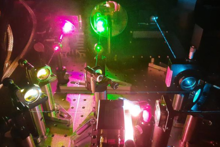
The new microscope uses optical fiber delivery of excitation light to enable very high image quality over a very large field of view with multicolor and high-speed capability. The researchers show that the instrument can be used to image liver cells, achieving a field of view up to 150 x 150 square micrometers and imaging rates up to 44 Hertz while maintaining a spatiotemporal resolution of less than 100 nanometers. “With this new microscope, individual drug combinations can be tested on isolated cells and then imaged with super-resolution to observe dynamics of cell membrane features or organelles,” said Ortkrass. “The large field of view can provide statistical information about the cell response, which could be used to improve personalized healthcare. Thanks to the system’s potentially small size, it might also be useful for clinical applications where high resolution is important.”
The new microscope is based on super-resolved structured illumination microscopy (SR-SIM), which uses a structured pattern of light to excite fluorescence in a sample and achieve a spatial resolution beyond the diffraction limit of light. SR-SIM is particularly well suited for live cell imaging because it uses low-power excitation that doesn’t harm the sample while producing highly detailed images. To achieve high resolution across a wide field of view, the new microscope reconstructs super-resolved images from a set of raw images. These raw images are acquired by using a set of six optical fibers to illuminate the sample with a sinusoidal striped pattern that is shifted and rotated to gain extra information. This creates a two-fold resolution improvement while still achieving fast imaging and being compatible with live-cell imaging.
“The fiber selection and phase shift is performed using a newly designed fiber switch based on galvanometric mirrors and MEMS-mirrors,” said Ortkrass. “We also custom-designed a hexagonal holder that collimates and refocuses the beams of the six fibers into the microscope to illuminate a large FOV and allow precise adjustment of all beams. This allows the setup to be used for total internal reflection fluorescence excitation (TIRF)-SIM, which is used to restrict fluorescence excitation and detection to a thin region of the sample.”
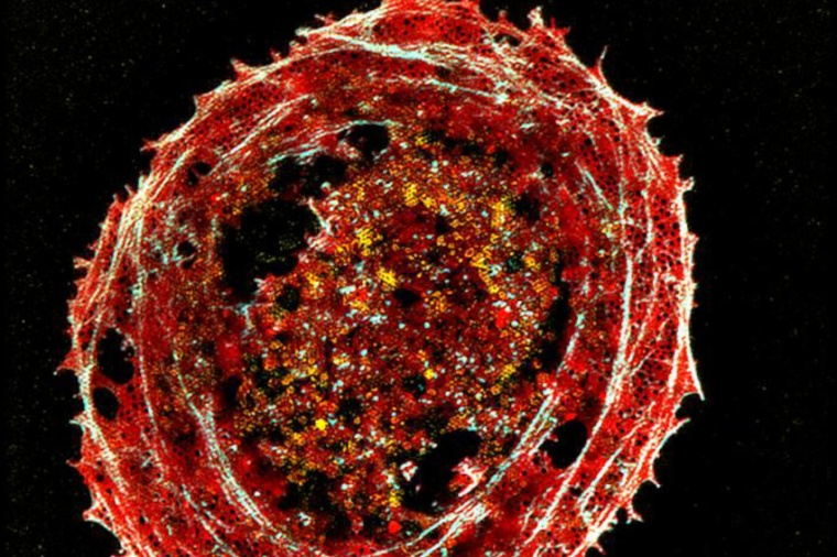
Since the liver is the primary organ involved in drug metabolism, the researchers tested the setup using samples of fixed multicolor-stained rat liver cells. The reconstructed images produced with the new microscope allowed visualization of the tiny membrane structures that are smaller than the diffraction limit of light. “This compact system uniquely combines a large field of view and fast pattern switching speed with multicolor, power-efficient excitation,” said Ortkrass. “In addition, the setup achieves very high image quality and can be tuned to perform either 2D-SIM or TIRF-SIM.”
Next, the researchers plan to apply the microscopy setup to live cell studies of liver cells to observe the dynamics of cells treated with several drugs. They also plan to improve the image reconstruction process to accomplish live reconstruction of the acquired raw data. (Source: Optica)
Link: Biomolecular Photonics Research Group, Faculty of Physics, Bielefeld University, Bielefeld, Germany


most read

New Jumo advertising tower with LED technology and a rotating logo in Fulda
The company has replaced the old tower at its Fulda site with a technically upgraded facility.
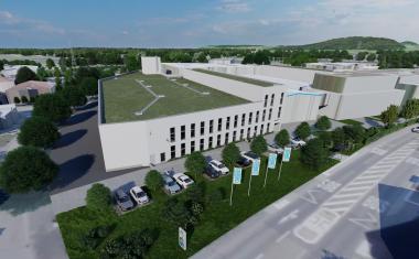
Hummel Expands Its Plant in Denzlingen with a Focus on Resource Efficiency
The company is planning a major plant expansion at its headquarters in Denzlingen and, following the completion of the planning phase, has submitted the building permit application.
Logimat 2027 Focuses on the Interaction Between People and Technology
Under the theme “Mind meets machine – what's next?”, the trade show focuses on the interplay between human expertise and technological systems.
Control 2027 Focuses on Modern Quality Assurance and AI-Powered Measurement Technology
Following the Control Expert Days 2026, the organizer, P. E. Schall, is preparing a program that reflects the latest developments in industrial quality assurance

Schaeffler bundles Defense and New Space activities under new CEO
Celia Pelaz takes over the position of CEO Defence & New Space at Schaeffler on October 1, 2026 and becomes Chairwoman of the Management Board of Stech Defence GmbH.