Supermicroscope shows nanoscale biological process for the first time
New device provides more insight into arterial calcification.
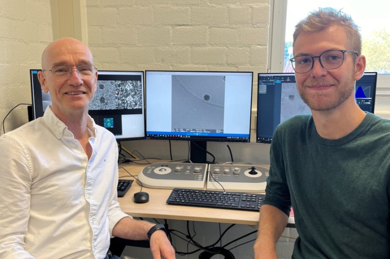
In Nijmegen, the world’s first microscope has been installed that is capable of live imaging of biological processes in such detail that moving protein complexes are visible. This new microscopic technique was developed by researchers led by Nico Sommerdijk from Radboud University Medical Center. As a demonstration of this innovative technique, Sommerdijk is now showcasing how arterial calcification begins. Previously, researchers faced a choice. They could use a microscope to examine material in detail down to the molecular level, but only in frozen, motionless samples. Alternatively, they could observe living, moving material, but with far less detail. Radboud researchers have developed a technique that combines both approaches.
This opens up various new possibilities for the future, such as visualizing how the COVID-19 vaccine enters a cell or capturing the initial stages of arterial calcification. There were many technical challenges. “If you want to see protein complexes in such fine detail, you need an electron microscope”, explains Nico Sommerdijk. “But the electron beam used can damage the biological material and the surrounding fluid, which is undesirable when you want to observe natural processes in the material over extended periods.” The solution is to apply a protective layer around the material to minimize damage from the electron beam. This can be achieved with graphene, an ultra-strong substance composed of a single layer of carbon atoms.
“But as soon as you apply it, the biological process you want to capture starts immediately”, Sommerdijk explains. “And then you have to quickly reach the microscope, locate the right spot in the tissue, and set up the microscope. This process takes at least half an hour, and sometimes the process is already over by then.” Sommerdijk and his team devised a method to address all these challenges. They apply a layer of graphene around the tissue and freeze it immediately, pausing biological processes. Then, using a light microscope, they identify the specific area in the tissue they want to visualize. Only after determining the correct orientation is the material placed in the new electron microscope, which can perform measurements in liquid. In this setup, the material is warmed, reactivating the biological processes, which are then directly visualized on a nanoscale.
As an example of what the new technique can reveal, Sommerdijk and his team now show how calcium deposits in a form that may lead to calcification of the arteries and aortic valve. PhD candidate Luco Rutten explains: “If there’s too much calcium phosphate in the blood, a particular protein in the body can bind to it, preventing it from precipitating. The kidneys then clear it out. Under the microscope, we see that these proteins form tiny spheres with calcium phosphate, which can still be broken down. But these spheres can also grow larger, causing calcium phosphate to turn into calcified deposits, which can no longer be broken down.” This may contribute to calcification in the body.
Currently, no treatment exists for calcified aortic valves other than complete valve replacement. “We still don’t fully understand what exactly happens with this type of calcification, which is why there are no medications yet”, says Sommerdijk. He aims to further study this with the new microscope and has recently received an ERC Advanced Grant to support this work. In this project, he plans to develop a “heart valve on a chip”. Initially, this will be a model of a healthy valve, into which he will then introduce calcification. This project will begin in 2025. (Source: Radboud UMC)