Switching off the light to see better
Switching fluorescent molecules on and off to create sharper microscope images of cell structures.
To study living organisms at ever smaller length scales, scientists must devise new techniques to overcome the diffraction limit. This is the intrinsic limitation on a microscope’s ability to focus on objects smaller than the wavelength of light being used. Structured illumination microscopy is one of the super-resolution techniques that can help, by illuminating sinusoidal-patterned light on a sample to obtain a sharper image. However, background light from out-of-focus regions can still smear out the final picture. Now, researchers from Osaka University demonstrated a new approach for super-resolution microscopy capable of observing structures inside a single cell or a cell cluster.
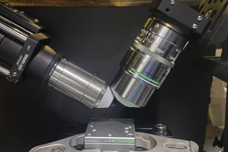
This was accomplished by selecting only a desired plane to image using thin light sheet illumination, projected perpendicular to the lens, to switch on fluorophores. “We show that selective-plane activation allows us to image dense microstructures inside cells with excellent sharpness not readily available previously,” Kenta Temma says. That is, sinusoidal structured light selectively excited only a thin plane where on-state fluorophores were localized, which allowed for background-free super-resolution imaging.
While some previous methods utilized random fluorescence emission from single molecules, or donut shaped second light source to deactivate or deplete fluorescent sources outside of a desired area, this new method can be gentler on cells that might be damaged by intense or long exposure to light. The researchers believe that their approach is especially effective when trying to understand what is happening in living systems with spatial structure, which can often exhibit background light outside the desired focal plane.
This includes organoids, which are artificial assemblies of different cell types meant to reproduce the behavior of actual body organs much better compared with collections of cells cultured on a flat petri dish. “We anticipate that our technique will be useful for future biological studies of 3D cell clusters, including organoids,” says Katsumasa Fujita. The same could apply to other complex biological systems. (Source: Osaka U.)
Link: Advanced Photonics and Biosensing Open Innovation Laboratory, AIST-Osaka University, Osaka, Japan