Wide high-speed imaging with super-resolution
12.09.2023 - Microscope based on fiberoptic components designed for studying cellular effects of drug combinations.
Researchers have developed a fluorescence microscope that uses structured illumination for fast super-resolution imaging over a wide field of view. The new microscope was designed to image multiple living cells simultaneously with a very high resolution to study the effects of various drugs and mixtures of drugs on the body. “Polypharmacy – the effect of the many combinations of drugs typically prescribed to the chronically sick or elderly – can lead to dangerous interactions and is becoming a major issue,” said Henning Ortkrass from Bielefeld University in Germany. “We developed this microscope to develop a platform that can investigate polypharmacy in individual patients.”
-
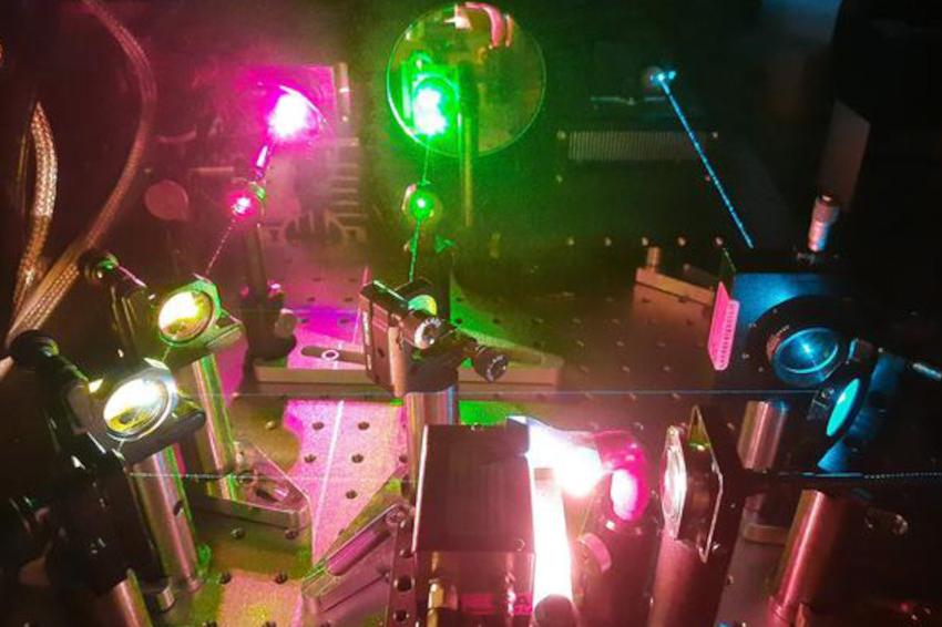 A fluorescence microscope uses structured illumination for fast super-resolution imaging over a wide field of view. It can also be used for multicolor and high-speed imaging. (Source: H. Ortkrass, Bielefeld Univ.)
 -
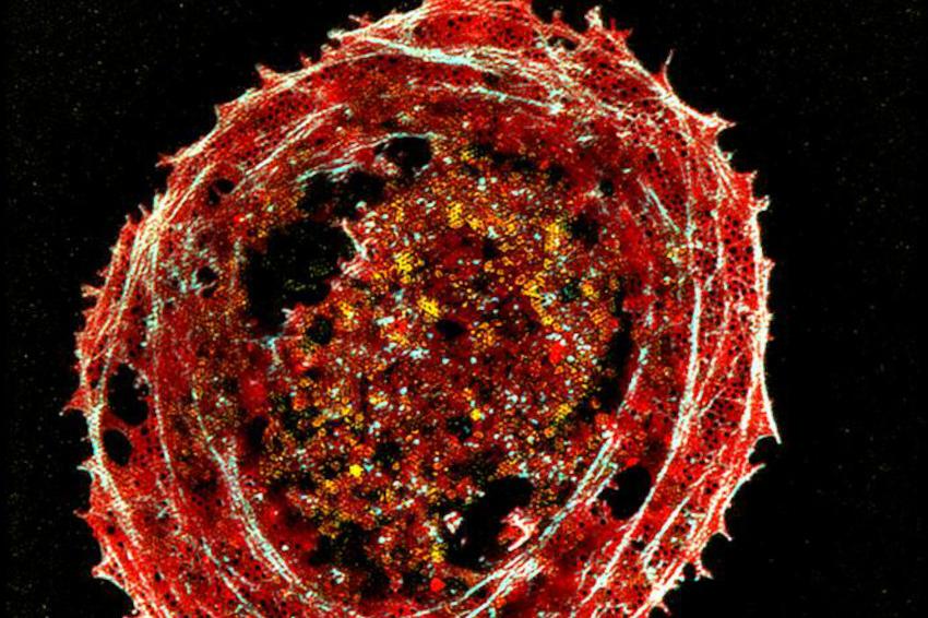 The image shows tiny membrane structures of fixed multicolor-stained liver cells, which are smaller than the diffraction limit of light. (Source: H. Ortkrass, Bielefeld Univ.)
The new microscope uses optical fiber delivery of excitation light to enable very high image quality over a very large field of view with multicolor and high-speed capability. The researchers show that the instrument can be used to image liver cells, achieving a field of view up to 150 x 150 square micrometers and imaging rates up to 44 Hertz while maintaining a spatiotemporal resolution of less than 100 nanometers. “With this new microscope, individual drug combinations can be tested on isolated cells and then imaged with super-resolution to observe dynamics of cell membrane features or organelles,” said Ortkrass. “The large field of view can provide statistical information about the cell response, which could be used to improve personalized healthcare. Thanks to the system’s potentially small size, it might also be useful for clinical applications where high resolution is important.”
The new microscope is based on super-resolved structured illumination microscopy (SR-SIM), which uses a structured pattern of light to excite fluorescence in a sample and achieve a spatial resolution beyond the diffraction limit of light. SR-SIM is particularly well suited for live cell imaging because it uses low-power excitation that doesn’t harm the sample while producing highly detailed images. To achieve high resolution across a wide field of view, the new microscope reconstructs super-resolved images from a set of raw images. These raw images are acquired by using a set of six optical fibers to illuminate the sample with a sinusoidal striped pattern that is shifted and rotated to gain extra information. This creates a two-fold resolution improvement while still achieving fast imaging and being compatible with live-cell imaging.
“The fiber selection and phase shift is performed using a newly designed fiber switch based on galvanometric mirrors and MEMS-mirrors,” said Ortkrass. “We also custom-designed a hexagonal holder that collimates and refocuses the beams of the six fibers into the microscope to illuminate a large FOV and allow precise adjustment of all beams. This allows the setup to be used for total internal reflection fluorescence excitation (TIRF)-SIM, which is used to restrict fluorescence excitation and detection to a thin region of the sample.”
Since the liver is the primary organ involved in drug metabolism, the researchers tested the setup using samples of fixed multicolor-stained rat liver cells. The reconstructed images produced with the new microscope allowed visualization of the tiny membrane structures that are smaller than the diffraction limit of light. “This compact system uniquely combines a large field of view and fast pattern switching speed with multicolor, power-efficient excitation,” said Ortkrass. “In addition, the setup achieves very high image quality and can be tuned to perform either 2D-SIM or TIRF-SIM.”
Next, the researchers plan to apply the microscopy setup to live cell studies of liver cells to observe the dynamics of cells treated with several drugs. They also plan to improve the image reconstruction process to accomplish live reconstruction of the acquired raw data. (Source: Optica)
Link: Biomolecular Photonics Research Group, Faculty of Physics, Bielefeld University, Bielefeld, Germany