Open multiplexing offers cancer researchers more choices
New features of Cell DIVE enable scalability for scientists with manual or automated workflows
The more scalable and efficient multiplexing platform addresses spatial cell biology and function within the tissue microenvironment, offering researchers the freedom to select from over 350 rigorously validated antibodies to design their studies in their own way and a proven workflow to image sixty-plus biomarkers in a single tissue section.
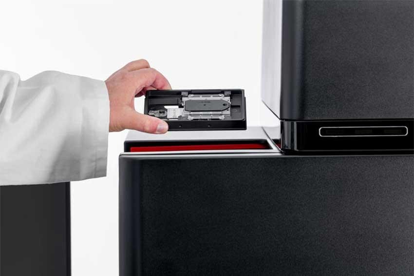
The new Cell DIVE ClickWell coverslip-free slide holder enables users to automate their workflows based on their unique needs. The improved Cell DIVE workflow reduces the risk of unnecessary tissue damage and improves sample processing time while still offering the researcher the freedom to choose the staining method that is best for their research.
“Cell DIVE enables scientists, particularly those working in cancer research, to map normal and diseased tissue by cell type, biomarker profile, and specific features better than ever before,” says James O’Brien, vice president of life sciences at Leica Microsystems. “Cell DIVE with ClickWell powers open multiplexing by making the manual workflow more efficient. It also gives researchers the power to choose their automation strategy to scale their research as it suits them.”
Cell DIVE sample staining occurs off the imager, allowing the user to process many slides through staining and dye inactivation steps in batches, thereby enabling scalability even without automation. ClickWell provides flexibility for automating sample staining, enabling the use of automated pipetting systems and autostainers. Researchers can also add a robotic arm to support unattended imaging of many samples.
Contact
Leica Microsystems GmbH
Ernst-Leitz-Str. 17-37
35578 Wetzlar
Germany
+49 6441 29 4000
+49 6441 29 4155


